Characterization of Extracellular Vesicles in Fibroblasts of Collagen-VI Related Muscular Dystrophy
A team led by the Applied research in neuromuscular diseases at the Institut de Recerca Sant Joan de Déu · SJD Barcelona Children's Hospital has made significant advancements in comprehending the pathogenesis of Collagen-VI Related Muscular Dystrophy (COL6-RD) by analyzing extracellular vesicles and their correlation with cellular mobility.
Dr. Cecilia Jiménez-Mallebrera, a researcher at IRSJD and CIBERER, led the team that, for the first time, conducted a proteomics analysis of the content of extracellular vesicles and the secretome in fibroblasts from patients with Collagen-VI Related Muscular Dystrophy (COL6-RD). These findings were published in the journal Scientific Reportsand involved collaboration with the Plataforma de Proteómica del Centro de Investigación Biomédica Navarrabiomed.
Currently, disorders related to collagen VI are the second most common congenital muscular dystrophies, for which there is no treatment. The severity of their manifestations ranges from comparatively mild Bethlem myopathy to significantly more severe Ullrich muscular dystrophies.
"Our objective was to investigate the significance of extracellular vesicles in intercellular communication as a potential mechanism implicated in the onset of the disease. Furthermore, we might identify new disease-related biomarkers by comparing the protein content of extracellular vesicles and the secreted proteins (secretome) released by fibroblasts from patients with COL6-RD with that of fibroblasts from unaffected children. These studies provide a comprehensive description of the affected cell in COL6-RD, which complements our previous transcriptomic findings. Commented Dr. Jiménez-Mallebrera, coordinator of the Applied research in neuromuscular diseases at IRSJD.
INTERESTING RESULTS FOR THE DEVELOPMENT OF NEW THERAPEUTIC STRATEGIES
"We have identified specific proteins that are associated with vesicle transport, actin cytoskeleton remodeling, cellular hemostasis, and oxidative stress that were detected in the extracellular vesicles of patients. These findings suggest that they could be involved in the pathogenesis of the disease." Commented Carmen Badosa (IRSJD), the first author of the article.
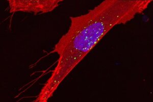
Indeed, in this study, we have demonstrated that the addition of extracellular vesicles derived from patient fibroblasts to control fibroblast cultures impacted the motility of the latter, resulting in modifications in speed and direction. To obtain these outcomes, we employed the wound healing assay and live cell imaging through microscopy, both of which were coordinated by Dr. Monica Roldán, who is the director of the Confocal Microscopy and Cellular Imaging Unit at SJD Barcelona Children's Hospital and an IRSJD researcher.
The in vitro wound healing method measures cell migration in a cell-confluent region by eliminating or excluding cells during culture. Subsequently, the closure of the wound is monitored through the utilization of in vivo microscopy imaging. This approach facilitates our comprehension of the movement of cells during biological processes, such as the spread of cancer, embryonic development, and tissue repair.
A video about the wound healing technique shows fibroblast cultures moving toward the wound to cover it. The first part of the video shows the cells, while the second part shows how to process images to extract information.
Furthermore, this study provides new insights into the role of extracellular vesicles in the pathogenesis of Collagen-VI Related Muscular Dystrophy. These patients experience difficulties with the healing and regeneration of various connective tissues, which may be attributed to our observations regarding cellular motility.
Dr. Cecilia Jiménez-Mallebrera, leader of the research team, concludes. "Our research presents the initial comprehensive account of extracellular vesicles in patients with congenital muscular dystrophy. It demonstrates that fibroblasts from these patients release a set of proteins that may reflect the diseases state and exert an impact on the function of the surrounding cells."
These findings present novel prospects for more precise diagnosis and treatment of Collagen-VI Related Muscular Dystrophy.
THE ROLE OF EXTRACELLULAR VESICLES IN CELL COMMUNICATION
The fundamental function of cell communication is to ensure the maintenance of tissue homeostasis, thereby ensuring their optimal and harmonious functioning within the body. This communication is imperative for the survival and proper functioning of all bodily systems. Some of this communication is achieved through the release of molecules into the extracellular space or through direct contact between cells. It is important to emphasize the crucial role of extracellular vesicles in the exchange of molecules, both short and long-range.
The extracellular vesicles are tiny structures released by cells, containing various components, including proteins, lipids, and nucleic acids. They can be transferred from one cell to another. Cell communication is dependent on these vesicles, and their examination yields valuable insights into the cells condition. Although their physiological function is yet to be fully comprehended, it is posited that they are implicated in numerous crucial processes, including but not limited to cell growth regulation, differentiation, immune response, and tissue repair. Moreover, it has been observed that they play a significant role in the etiology of diverse ailments.
Reference
Badosa C, Roldán M, Fernández-Irigoyen J, Santamaria E, Jimenez-Mallebrera C. Proteomic and functional characterisation of extracellular vesicles from collagen VI deficient human fibroblasts reveals a role in cell motility. Sci Rep. 2023 Sep 5;13(1):14622. doi: 10.1038/s41598-023-41632-1. PMID: 37670049; PMCID: PMC10480450.
Our research provides the first detailed description of extracellular vesicles in patients with congenital muscular dystrophy, and opens new opportunities for better diagnosis and treatment.
