Gene therapy proved against Muscular Distrophy with the ALBA Synchrotron
A study by the Institut de Recerca Sant Joan de Déu, ICFO, CIBERER and the ALBA Synchrotron has helped demonstrate that gene therapy can reverse the effects of the mutation that causes the symptoms of congenital muscular dystrophy in patient cells. The mutation, which leads to a disorder in the body's collagen, has been silenced through a genetic editing technique based on the CRISPR/Cas9 system.
Congenital muscular dystrophy is a group of rare neuromuscular diseases. In particular, type VI collagen deficiency-related dystrophy affects less than 1 in 100,000 people, has varying degrees of severity, and has no cure.
Collagen VI is present in a large number of tissues in our body: muscles, tendons, cartilage, blood vessels or even the brain. The cells that produce it, called fibroblasts, secrete it out into the space between cells, and the collagen fibers create a network whose function is to maintain the integrity and function of said body tissues. That is why a deficiency in this collagen VI, such as that suffered by people with muscular dystrophy, leads to high level of disability and low life expectancy.
Now, a study led by the Institut de Recerca Sant Joan de Déu (IRSJD) and the Institute of Photonic Sciences (ICFO), within the ICFO-SJD JointLab program, with the participation of the Center for Biomedical Network Research on Rare Diseases (CIBERER) and the ALBA Synchrotron has shown promising results with gene therapy to combat muscular dystrophy. The analysis of patient's cells has made it possible to see the alterations suffered due to the disease and has proven how the experimental treatment with gene therapy helps to reverse these defects.
Patients with muscular dystrophy have a mutation in the genes that are responsible for producing collagen, so that they cannot correctly form this network of collagen fibers between cells. The objective of the scientific team was to silence this mutation with a genetic editing technique based on the CRISPR/Cas system.
For the study, they took skin biopsies from several children diagnosed with muscular dystrophy. Once they had these fibroblast cells, they used gene therapy in the lab to more or less correctly rewrite these collagen-coding genes. The results are very satisfactory since they have achieved that the patient's cells once again have the appearance of healthy cells.
New discoveries under the synchrotron light
Verify the effectiveness of the gene therapy treatment, the research team has analyzed the cells of the patients with advanced microscopy techniques. Among them, the super-resolution microscopy carried out in the Confocal Microscopy and Cell Imaging platform of the IRSJD and ICFO and the X-ray microscopy with synchrotron light in the MISTRAL beamline of ALBA. The joint work between these platforms has been key to understanding the results of the experiments.
"Thanks to the collaboration between our institution and ALBA, cells from patients with muscular dystrophy have been analyzed for the first time with synchrotron light. This collaboration has allowed us to take a step forward in the study of the disease and the search for possible treatments", explains Mónica Roldan, IRSJD researcher.
MISTRAL is a very unique microscope, as there are only 3 like it in the world in three other synchrotrons. "It allows doing tomography on the cells, like a hospital CT scan, but with a million times more resolution," explains Ana Joaquina Pérez Berná, a scientist at ALBA. This allows us to see the inside of the cells and understand what happens in them globally, since it is not necessary to cut or treat them chemically, they are only frozen.
The researchers also viewed the samples under the high-resolution STED microscope at ICFO. "This technique allows us to see the intracellular organelles much better, with higher resolution, and to carry out experiments that could not be done before," explains Enrico Castroflorio, postdoctoral researcher of the ICFO SLN team led by Pablo Loza-Alvarez. The group has long been developing microscopes and photonic technologies potentially useful as diagnostic tools.
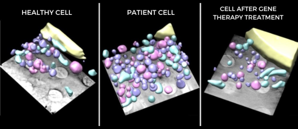
Analyzing the images obtained, the research team was able to observe that the cells of the patients had abnormalities inside them that were not expected. In addition to defects in the extracellular collagen matrix, they discovered an accumulation of intracellular organelles, such as endosomes and lysosomes. "This suggests that the collagen derived from the mutation also deregulates the intracellular space of the fibroblasts in patients with muscular dystrophy", comments Pérez Berná.
The images of healthy fibroblast cells were compared with the images of cells affected by the mutation and with the cells treated with gene therapy, and showed how, effectively, the disorders derived from the mutation in the genes had been corrected. Their effects subsided and the normal collagen network between cells, as well as the levels of endosomes and lysosomes, were restored.
The IRSJD researcher, Cecilia Jiménez-Mallebrera, celebrates that "this is a great result since it confirms at the subcellular level how gene editing is capable of reversing not only the expression of the mutated gene at the RNA and protein level, but also at the functional. With other more standard techniques we could not have reached the same level of detail. This is due to the fact that there is a degree of uncertainty due to the possible artifacts generated in the preparation of the samples that does not exist with the technique used in ALBA. These results give us confidence to take the next step and test this therapy in mice."
Reference paper
Castroflorio, E.; Pérez Berná, A.J.; López-Márquez, A.; Badosa, C.; Loza-Alvarez, P.; Roldán, M.; Jiménez-Mallebrera, C. The Capillary Morphogenesis Gene 2 Triggers the Intracellular Hallmarks of Collagen VI-Related Muscular Dystrophy. Int. J. Mol. Sci. 2022, 23, 7651. https://doi.org/10.3390/ijms23147651

Experiments at the MISTRAL beamline in ALBA have revealed previously unknown cell damage.Congenital muscular dystrophy is a rare minority disease that mainly affects children and has no treatment.
