Diagnostic and Therapeutic Imaging
Research Program
Leaders
Where we are
 SJD Barcelona Children's Hospital
Related websites
The main thrust of the research of the Diagnostic and Therapeutic Imaging group is patient-oriented. The primary objectives are: 1) to identify imaging diagnostic or prognostic biomarkers, and 2) to develop and implement new image-based technologies. The latter includes the development of new techniques and improvements in the detection of injuries (artificial intelligence), safety and quality (radiation protection, interventional radiology) or innovative methodologies (development of own calibration models or virtual or printed 3D segmentation).
In addition to investigating these different lines of research, we also offer support to scientific projects of other IRSJD or external groups relating, for example, to imaging design, acquisition and analysis.
Research lines
- Vascular disease, focused mainly on examining the progression of morpho-functional aspects as well as the identification of new prognostic biomarkers of neonatal cerebrovascular injury.
- Hearing defects, aimed at identifying new biomarkers and elucidating the responsible structural bases.
- Biomarkers of sarcoma treatment response, with the validation of new biomarkers and multiparametric techniques.
- Development of tools and procedures for the optimisation of imaging techniques.
Scientific objectives
- To develop standardised population models for different imaging markers by implementing post-process tools that would allow patients to be compared against standardised models (morphometry techniques, connectivity and tissue characterisation, etc.).
- To develop and validate paediatric vascular imaging biomarkers for population-based disease-progression studies, as well as for the diagnosis and monitoring of diseases of possible cerebrovascular origin.
- To develop biomarkers based on diffusion techniques for soft tissue solid tumours (sarcomas).
- To develop biomarkers for the study of imaging factors involved in both childhood developmental and hearing disorders.
- To develop prognostic imaging indicators of developmental impairment and neurodegenerative diseases.
- To develop a radiological decision support tool based on artificial intelligence algorithms for the detection and classification of different types of lesions on a basic chest x-ray.
- To develop strategies to minimise anaesthesia and acquisition time in young paediatric patients, and patients with behavioural disorders or with medical implants.
Area/Field of expertise
The research carried out by our group is part of the study and applicability of diagnostic imaging techniques (basic or contrast radiology, ultrasound, magnetic resonance imaging [MRI], computed tomography [CT] or their combinations with nuclear medicine). We would like to draw special attention to advanced techniques (multiparametric MRI, diffusion-tensor imaging, functional MRI) and the development of new imaging areas of research (3D planning, radiation dose management, artificial intelligence, etc.).
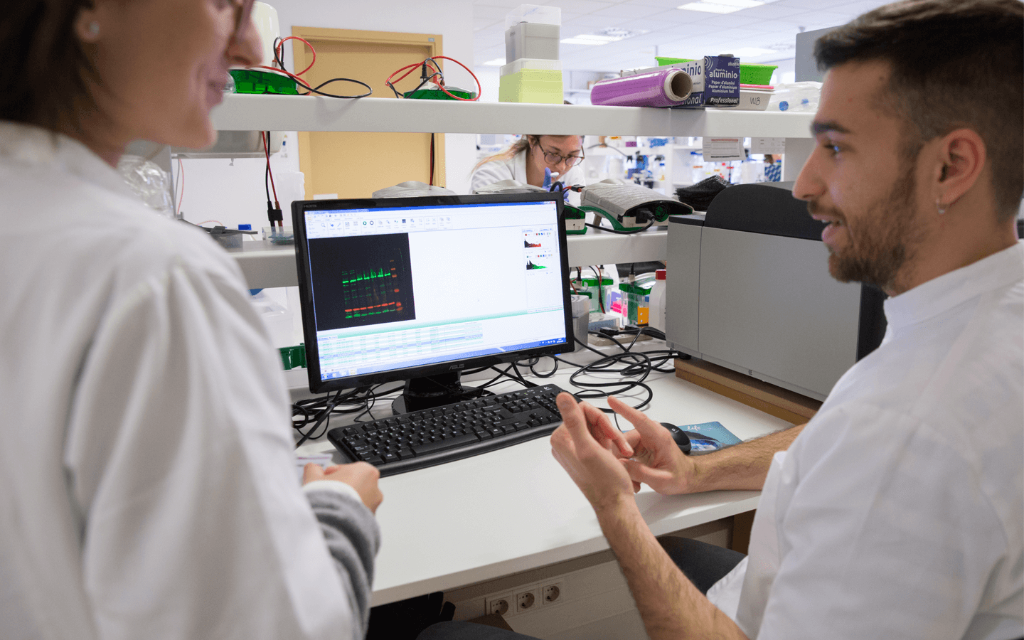
We are a group of radiologists and imaging technicians who provide a comprehensive approach to the methods used and the data that can be extracted using different imaging techniques.
At present our department stands out for the quality of its technology: two 1.5 Tesla (General Electric) and one 3 Tesla (Philips) MRIs, a multi-slice CT scanner with 256-row detector and iterative image acquisition programmes (linked to programmes to detect and analyse the amount of radiation absorbed) as well as two high-performance ultrasound scanners (one with elastography imaging). We also have technology for functional studies (presentation of audiovisual stimuli and manual response) in MRI as well as various post-processing programmes using the Philips Intellispace Portal solution.
Over the past year our department has conducted over 79,000 studies, approximately 7,600 MRIs, 11,000 ultrasound scans and 4,000 CT scans. We participated in 40 clinical trials here in the Hospital and several research projects by external researchers, including approximately 250 scans in the 3 Tesla MRI.
Group members
-

Jefe de Grupo
-
Ignasi Barber Martínez De La Torre
Investigador
-
Investigador
-
Investigador
-
Investigador post-doc
-
Investigador
-
Jaime Monturiol Duran
Investigador
Last Publications
- Inarejos E, Navallas M, Enrique Ladera Gonzalez, Gómez-Chiari M, Torner-Rubies F and Navarro OM Pediatric vertebral tumors: radiologic features and differential diagnoses. PEDIATRIC RADIOLOGY . 56(2): 297-313.
- Dias, SC, Habre, C, Di Paolo, PL, d'Angelo, P, Augdal, TA, Angenete, OW, Kljucevsek, D, Inarejos E, de Horatio, LT and Rosendahl, K ESR Essentials: juvenile idiopathic arthritis; what every radiologist needs to know-practice recommendations by the European Society of Paediatric Radiology EUROPEAN RADIOLOGY . 36(2): 1261-1271.
- Nievelstein, RAJ, Borgwardt, L, Inarejos E, Von Kalle, T, Kyncl, M, Lequin, MH, Littooij, AS, Pace, E, Di Paolo, PL, Van Rijn, RR, Rogasch, JMM, Schäfer, J, Pasic, IS, Spezzacatene, A, Tolboom, N, Wan, SM and Zucchetta, P White paper on the current state of imaging biomarkers in paediatric oncology PEDIATRIC RADIOLOGY . 55(12): 2493-2507.
Projects
- Project name:
- Radiation, Health, Safety and Quality for Youth: A Comprehensive Approach to Justification, Optimisation, and Education
- Leader
- Ignasi Barber Martínez de la Torre
- Funding entities:
- European Commission
- Code
- 101232948
- Starting - finishing date:
- 2025 - 2029
- Project name:
- Artificial intelligence (AI) based - CArdiovascular Risk in ONcological pediatric patients assessment Tools
- Leader
- Jose Luis Munuera del Cerro, Arnau Valls Esteve
- Funding entities:
- Ministerio De Ciencia E Innovacion
- Code
- TED2021-132025B-C44
- Starting - finishing date:
- 2022 - 2025
- Project name:
- Investigo 2022_Josep Munuera
- Leader
- Jose Luis Munuera del Cerro
- Funding entities:
- Agaur - Agència de Gestió d'Ajuts Universitaris i de Recerca
- Code
- 100031TG8
- Starting - finishing date:
- 2022 - 2024
