
We identify a new cellular mechanism involved in Charcot-Marie-Tooth disease
A research team from the Neurogenetics and Molecular Medicine group of the Institut de Recerca Sant Joan de Déu (IRSJD), has identified a new molecular mechanism that helps explain the origin of Charcot-Marie-Tooth disease caused by mutations in the GDAP1 gene, one of the most common hereditary neuropathies. The study GDAP1 orchestrates redox signaling at membrane contact sites to preserve axonal integrity in Charcot-Marie-Tooth disease, published in Neurobiology of Disease, shows that the mitochondrial protein GDAP1 is essential for different structures inside nerve cells to work in a coordinated manner. When this protein does not function correctly, this communication is altered and, over time, axons -the extensions that transmit nerve impulses- deteriorate. The research involved the Confocal Microscopy Unit and collaborators from the Medical College of Wisconsin.
Charcot-Marie-Tooth
Charcot-Marie-Tooth disease (CMT) is a group of rare, inherited diseases that affect the peripheral nerves, which are responsible for transmitting signals between the brain, spinal cord, and the rest of the body. As these nerves deteriorate, affected individuals may experience muscle weakness and atrophy, difficulty walking, balance problems, and a progressive loss of sensation, especially in the feet and hands. Although there is currently no cure, research is key to understanding the mechanisms that cause the disease and developing new treatments that can slow its progression.
When the structures of the cell stop working in coordination
To function properly, neurons need the different structures inside them to work in a coordinated manner and communicate with each other. In this study, researchers have discovered that the GDAP1 protein plays an essential role in this coordination, as it helps maintain communication between the structures responsible for producing energy, obtaining nutrients and protecting the cell from damage caused by oxidative stress.
Analyzing cells from patients with Charcot-Marie-Tooth disease and an experimental mouse model, the team observed that, when the GDAP1 protein does not work properly, this communication between organelles is broken. As a result, the cells lose the ability to maintain their internal balance, the metabolism of fats necessary for the proper functioning of nerves and axons is altered, the extensions that transmit nerve impulses, and they end up deteriorating progressively.
When nerves begin to deteriorate
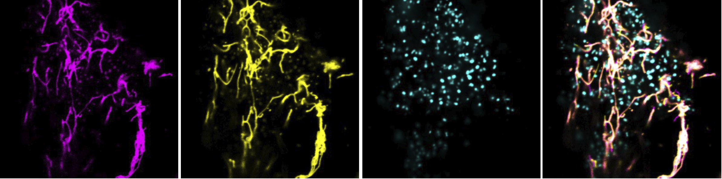
The research team also demonstrated that these alterations within the cells end up directly affecting the structure of the nerves. In the mouse model studied, they observed that several components essential for the proper functioning of neurons were no longer distributed correctly and that some of the structures responsible for transmitting nerve impulses quickly and efficiently, such as the node of Ranvier, deteriorated.
In addition, the cells showed an increase in oxidative stress-an imbalance that can cause cell damage-and alterations in processes essential for maintaining nerve health. Taken together, the results indicate that the lack of the GDAP1 protein triggers a series of alterations that, over time, favor the degeneration of axons, the nerve fibers responsible for transmitting information between the brain, spinal cord and the rest of the body.
New opportunities for treatment development
One of the most promising results of the study is that the researchers were able to partially restore normal cell function in experimental models. They did this by both restoring the function of the GDAP1 protein and correcting the chemical imbalance that causes its alteration.
Although these results are still in the early stages of research, they indicate that these mechanisms could become a new avenue for developing treatments capable of slowing the progression of Charcot-Marie-Tooth disease in the future. This focus is part of the collaboration that the group maintains with IBEC as part of a project funded by Torrons Vicencs-RAC1.
Researcher Lara Cantarero from the Neurogenetics and Molecular Medicine research group at the IRSJD and first author of the study comments: "We knew that the GDAP1 protein was important for the functioning of nerve cells, but now we have discovered that it is also essential for the different structures of the cell to work in a coordinated manner. When this communication is broken, the nerves begin to deteriorate."

One of the most promising results of the study is that the researchers were able to partially restore normal cell function in experimental models.
